Patient Education: Adult Hip Dysplasia
Education
Hip Dysplasia
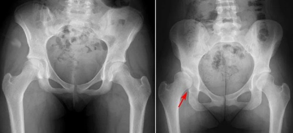
Hip dysplasia is a condition where the hip socket (acetabulum) is too shallow to fully cover and support the head of the femur (thigh bone). This lack of coverage can lead to increased stress on the cartilage and labrum, resulting in pain, instability, and an increased risk of arthritis over time.
Hip dysplasia is one of the most common structural causes of early-onset hip pain and arthritis, especially in young adults.
What is Hip Dysplasia?
In a normal hip, the socket provides deep coverage of the femoral head, allowing stable movement while distributing forces evenly across the joint. In hip dysplasia, the socket is underdeveloped or shallow, leading to:
Reduced coverage of the femoral head
Increased stress on the labrum and cartilage
Greater risk of joint instability and cartilage breakdown
The degree of dysplasia can vary, from mild undercoverage to severe deformities that cause early joint damage.
What Causes Hip Dysplasia?
Hip dysplasia often develops during early growth and development. Some known factors include:
Genetics: A family history of hip dysplasia increases the risk.
Breech position at birth: Babies born in breech position have a higher risk.
Female sex: Hip dysplasia is more common in women.
Ligamentous laxity: Increased joint flexibility can contribute to instability.
While some cases are diagnosed in infancy, milder forms of dysplasia often go unrecognized until adulthood, when symptoms begin to develop.
Symptoms of Hip Dysplasia
Symptoms of hip dysplasia can vary but often include:
Pain in the front (groin) or side of the hip
Clicking, catching, or locking sensations
Feeling of instability or giving way
Stiffness, especially after sitting or activity
Progressive discomfort with sports or prolonged activity
Without treatment, dysplasia can lead to early cartilage wear and arthritis.
Diagnosis
Diagnosis involves:
History and physical exam: Evaluating hip motion, strength, and stability.
X-rays: To assess the depth of the socket and coverage of the femoral head.
Advanced imaging (MRI or CT): Used to evaluate cartilage, labral damage, and to assess three-dimensional hip anatomy.
Identifying hip dysplasia early is important to prevent or slow joint degeneration.
Treatment Options
Treatment depends on the severity of dysplasia and patient symptoms.
Non-Surgical Treatment:
Activity modification to reduce joint stress
Physical therapy to strengthen surrounding muscles and improve mechanics
Anti-inflammatory medications or injections for symptom relief
These options may help manage mild dysplasia, but they do not correct the underlying structural problem.
Surgical Treatment:
For patients with significant dysplasia and symptoms, surgery may be recommended to restore normal hip alignment and mechanics.
The most common procedure is the Periacetabular Osteotomy (PAO), which repositions the socket to improve femoral head coverage and distribute load more evenly.
If cartilage or labral damage is present, additional procedures such as labral repair or cartilage restoration may be performed at the same time.
The goal of surgery is to relieve pain, restore function, and preserve the natural hip joint.
Recovery and Outlook
Non-Surgical Management:
Focuses on symptom relief and protecting the joint but may not prevent progression in more severe cases.Surgical Recovery:
After PAO or related procedures:Protected weight-bearing initially
Gradual rehabilitation to restore motion and strength
Return to higher-impact activities usually around 6–12 months, depending on healing
Early surgical intervention in the right patients can improve symptoms and delay or prevent arthritis progression.
Conclusion
Hip dysplasia is a common cause of hip pain and dysfunction in young adults. Early recognition and appropriate treatment can improve pain, restore function, and protect the joint from long-term damage.
If you’re experiencing hip pain, instability, or limited activity tolerance, contact our office for a complete evaluation and personalized treatment plan.